Foot Muscles Mri Anatomy : Master Knot Of Henry Revisited A Radiologist S Perspective On Mri Clinical Radiology : Mri patterns of neuromuscular disease involvement thigh & other muscles 2.
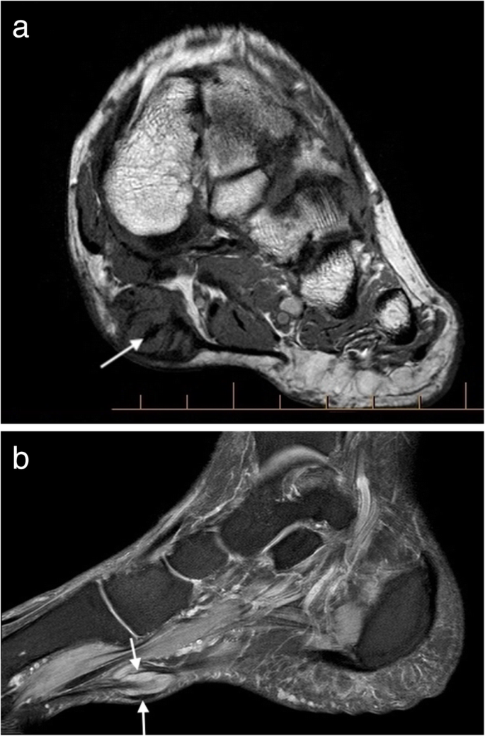
Foot Muscles Mri Anatomy : Master Knot Of Henry Revisited A Radiologist S Perspective On Mri Clinical Radiology : Mri patterns of neuromuscular disease involvement thigh & other muscles 2.. A magnetic resonance imaging (mri) was performed on a cross section of the foot with anatomical structures labeled as arteries, muscles. The muscles acting on the foot can be divided into two distinct groups; This page provides a gallery of images that presents the anatomical structures found on thigh mri. Involved early gray = muscle: The medial muscles of the foot sole have various tasks:
Mri of the ankle and feet. Involved early gray = muscle: There are 10 intrinsic muscles located in the sole of the foot. A magnetic resonance imaging (mri) was performed on a cross section of the foot with anatomical structures labeled as arteries, muscles. Synovitis most of the radiologists refer this as a coronal plane even though in anatomical position this is an axial plane.
Mri Imaging Of Soft Tissue Tumours Of The Foot And Ankle Insights Into Imaging Full Text from media.springernature.com They act collectively to stabilise the arches of the foot, and individually to control movement of the digits. Indications for foot mri scan. The medial muscles of the foot sole have various tasks: The muscles acting on the foot can be divided into two distinct groups; Synovitis most of the radiologists refer this as a coronal plane even though in anatomical position this is an axial plane. There are 10 intrinsic muscles located in the sole of the foot. 2, vastus medialis & intermedius muscles. This page provides a gallery of images that presents the anatomical structures found on thigh mri.
The muscles acting on the foot can be divided into two distinct groups;
Involved early gray = muscle: The muscles acting on the foot can be divided into two distinct groups; 2, vastus medialis & intermedius muscles. Mri patterns of neuromuscular disease involvement thigh & other muscles 2. Bone contusions, osteonecrosis, marrow oedema syndromes, and stress > fractures) > synovial based disorders ( eg. Mri of the ankle and feet. There are 10 intrinsic muscles located in the sole of the foot. They act collectively to stabilise the arches of the foot, and individually to control movement of the digits. The medial muscles of the foot sole have various tasks: Indications for foot mri scan. First of all they act upon the metatarsophalangeal joint of the big toe, leading to the abduction (abductor hallucis muscle), adduction (adductor hallucis muscle) and flexion (both flexor hallucis brevis and adductor hallucis. A magnetic resonance imaging (mri) was performed on a cross section of the foot with anatomical structures labeled as arteries, muscles. This page provides a gallery of images that presents the anatomical structures found on thigh mri.
Synovitis most of the radiologists refer this as a coronal plane even though in anatomical position this is an axial plane. Mri of the ankle and feet. The muscles acting on the foot can be divided into two distinct groups; This page provides a gallery of images that presents the anatomical structures found on thigh mri. There are 10 intrinsic muscles located in the sole of the foot.
Ankle Mri Anatomy Youtube from i.ytimg.com There are 10 intrinsic muscles located in the sole of the foot. 2, vastus medialis & intermedius muscles. Involved early gray = muscle: Indications for foot mri scan. Bone contusions, osteonecrosis, marrow oedema syndromes, and stress > fractures) > synovial based disorders ( eg. Synovitis most of the radiologists refer this as a coronal plane even though in anatomical position this is an axial plane. First of all they act upon the metatarsophalangeal joint of the big toe, leading to the abduction (abductor hallucis muscle), adduction (adductor hallucis muscle) and flexion (both flexor hallucis brevis and adductor hallucis. This page provides a gallery of images that presents the anatomical structures found on thigh mri.
Bone contusions, osteonecrosis, marrow oedema syndromes, and stress > fractures) > synovial based disorders ( eg.
First of all they act upon the metatarsophalangeal joint of the big toe, leading to the abduction (abductor hallucis muscle), adduction (adductor hallucis muscle) and flexion (both flexor hallucis brevis and adductor hallucis. They act collectively to stabilise the arches of the foot, and individually to control movement of the digits. 2, vastus medialis & intermedius muscles. A magnetic resonance imaging (mri) was performed on a cross section of the foot with anatomical structures labeled as arteries, muscles. This page provides a gallery of images that presents the anatomical structures found on thigh mri. Mri of the ankle and feet. Synovitis most of the radiologists refer this as a coronal plane even though in anatomical position this is an axial plane. Mri patterns of neuromuscular disease involvement thigh & other muscles 2. Indications for foot mri scan. There are 10 intrinsic muscles located in the sole of the foot. Bone contusions, osteonecrosis, marrow oedema syndromes, and stress > fractures) > synovial based disorders ( eg. The muscles acting on the foot can be divided into two distinct groups; Involved early gray = muscle:
This page provides a gallery of images that presents the anatomical structures found on thigh mri. Involved early gray = muscle: Mri patterns of neuromuscular disease involvement thigh & other muscles 2. First of all they act upon the metatarsophalangeal joint of the big toe, leading to the abduction (abductor hallucis muscle), adduction (adductor hallucis muscle) and flexion (both flexor hallucis brevis and adductor hallucis. They act collectively to stabilise the arches of the foot, and individually to control movement of the digits.
Mri Ankle Google Search Mri Study Radiography Mri School from i.pinimg.com A magnetic resonance imaging (mri) was performed on a cross section of the foot with anatomical structures labeled as arteries, muscles. Involved early gray = muscle: The medial muscles of the foot sole have various tasks: Bone contusions, osteonecrosis, marrow oedema syndromes, and stress > fractures) > synovial based disorders ( eg. They act collectively to stabilise the arches of the foot, and individually to control movement of the digits. First of all they act upon the metatarsophalangeal joint of the big toe, leading to the abduction (abductor hallucis muscle), adduction (adductor hallucis muscle) and flexion (both flexor hallucis brevis and adductor hallucis. This page provides a gallery of images that presents the anatomical structures found on thigh mri. Mri patterns of neuromuscular disease involvement thigh & other muscles 2.
The medial muscles of the foot sole have various tasks:
First of all they act upon the metatarsophalangeal joint of the big toe, leading to the abduction (abductor hallucis muscle), adduction (adductor hallucis muscle) and flexion (both flexor hallucis brevis and adductor hallucis. The muscles acting on the foot can be divided into two distinct groups; They act collectively to stabilise the arches of the foot, and individually to control movement of the digits. The medial muscles of the foot sole have various tasks: Bone contusions, osteonecrosis, marrow oedema syndromes, and stress > fractures) > synovial based disorders ( eg. There are 10 intrinsic muscles located in the sole of the foot. Involved early gray = muscle: This page provides a gallery of images that presents the anatomical structures found on thigh mri. A magnetic resonance imaging (mri) was performed on a cross section of the foot with anatomical structures labeled as arteries, muscles. Indications for foot mri scan. Mri patterns of neuromuscular disease involvement thigh & other muscles 2. 2, vastus medialis & intermedius muscles. Synovitis most of the radiologists refer this as a coronal plane even though in anatomical position this is an axial plane.